
|
||||||||||||||||||||||
|
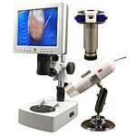
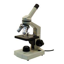
Microscopes Here you can see temperature meters from these companies:
Technical specifications for our Microscopes can be found at the following links: |
||||||||||||||||||||||
|
- PCE-MM 200 Microscopes |
 |
|||||||||||||||||||||
|
- MikroCam
Microsopes |
 |
|||||||||||||||||||||
|
- 5283000
Microscopes |
 |
|||||||||||||||||||||
|
- Microscopes
DigiMicro 2.0 Deluxe |
 |
|||||||||||||||||||||
|
- PCE-MM
200UV Microscopes |
 |
|||||||||||||||||||||
|
- XDS-2
FL Microscopes |
 |
|||||||||||||||||||||
|
- Microscopes
DigiMicro 2.0 Scale |
 |
|||||||||||||||||||||
|
- 52-81000
Microscopes |
 |
|||||||||||||||||||||
|
- XZ-2
Microscopes |
|
|||||||||||||||||||||
|
- Microscopes
DigiMicro Profi |
 |
|||||||||||||||||||||
|
- Microscopes
B-353FL |
 |
|||||||||||||||||||||
|
- Microscopes
DigiMicro Labs5.0 |
 |
|||||||||||||||||||||
|
- Microscopes
DigiMicro Mobile |
 |
|||||||||||||||||||||
|
-
Microscopes PCE-IVM 3D |
 |
|||||||||||||||||||||
|
- Microscopes
PCE-VM 21 |
 |
|||||||||||||||||||||
|
- Microscopes
PCE-MVM 3D |
 |
|||||||||||||||||||||
|
-
Microscopes
BioDiscover |
 |
|||||||||||||||||||||
|
-
Microscopes
XDS-3FL |
 |
|||||||||||||||||||||
|
-
Microscopes
Duolux |
 |
|||||||||||||||||||||
|
-
Microscopes
MML1200 |
 |
|||||||||||||||||||||
|
-
Microscopes
Erudit DLX |
 |
|||||||||||||||||||||
|
-
Microscopes
MBL3400 |
 |
|||||||||||||||||||||
|
-
Microscopes
XDS-3MET |
 |
|||||||||||||||||||||
|
-
Microscopes
Erudit MO |
 |
|||||||||||||||||||||
|
-
Microscopes
Biolux ICD |
 |
|||||||||||||||||||||
|
-
Microscopes
MSL4000-10/30-IL-TL |
 |
|||||||||||||||||||||
|
-
Microscopes
MSL4000-20/40-IL-TL |
 |
|||||||||||||||||||||
|
-
Microscopes
Biorit ICD |
 |
|||||||||||||||||||||
|
-
Microscopes
Biorit ICD-CS |
 |
|||||||||||||||||||||
|
-
Microscopes
MSZ5000 |
 |
|||||||||||||||||||||
|
-
Microscopes
MSZ5000-IL-TL |
 |
|||||||||||||||||||||
|
-
Microscopes
B-353LD |
|
|||||||||||||||||||||
|
-
Microscopes
MSZ5000-T-IL-TL |
 |
|||||||||||||||||||||
|
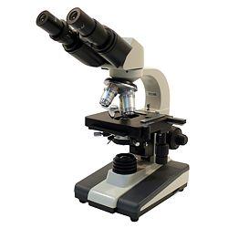
- Bino
Researcher Microscopes |
 |
|||||||||||||||||||||
|
- Microscopes
XC-100L |
 |
|||||||||||||||||||||
|
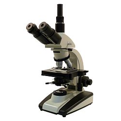
- Trino
Researcher Microscopes |
 |
|||||||||||||||||||||
|
- Advance
ICD 10x-160x Microscopes |
 |
|||||||||||||||||||||
|
-
Microscopes
Science IVM-401 |
 |
|||||||||||||||||||||
|
-
Microscopes
Science MPO-401 |
 |
|||||||||||||||||||||
|
-
Microscopes
Science ADL-601F |
 |
|||||||||||||||||||||
|
-
Microscopes
B-600TiFl |
 |
|||||||||||||||||||||
|
- Microtome
MT.5503 |
 |
|||||||||||||||||||||
|
-
Microtome MT.5505 |
 |
|||||||||||||||||||||
|
- Microtome MT.5501 |
 |
|||||||||||||||||||||
|
- Endoscopes |
|
|||||||||||||||||||||
|
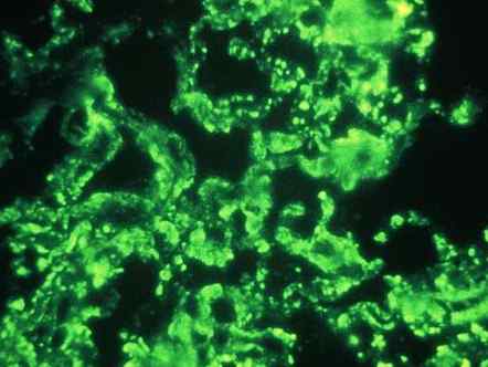
The areas of use for microscopes is nealry unlimited.
|
||||||||||||||||||||||


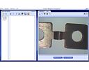
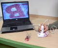
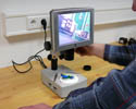
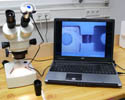
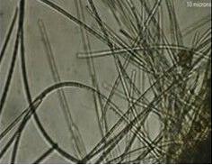 es of small items. Those icroscopes increase the
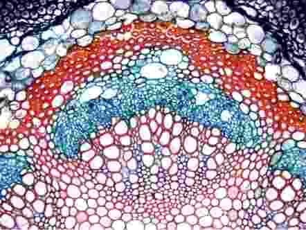

|
You are currently at: Home / measuring instruments and test equipment / Microscopes |
|
If you have any questions, call our offices on: |
|
This page in German |
