|
Microscopes Microscopes are composed of a magnifying lens and an eyepiece that join together in a tube, an illuminated stage where the object being observed is placed and a tripod to secure the optical components. The source of light for our microscopes is generally a small bulb attached to the tripod of lenses that can be adjusted (via a lens or system of lenses near the bulb) and fixed behind the illuminated area. The condenser for our microscopes is a complicated system of lenses or mirrors that reproduce the light on the surface of the object. The lens for our microscopes amplifies the object so that this magnified image can be viewed in the eyepiece. Microscopes also have two eyepieces to allow the object to be viewed with both eyes. This distinguishes a microscope using transmitted light that passes through very thin transparent objects, and microscopes using reflected light for analysing surfaces of opaque objects. We offer a range of microscopes for various purposes, such as for use in laboratories, research, in the office or for a hobby.
There are even microscopes for the application in schools. Since young students
do also face microscopy in class, microscopes with high accuracy are necessary
to guarantee an optimal education fun. All of these microscopes can be connected via a USB port to a computer making it possible to document images
or record videos for analysis or even to a projector to display the images to a wider audience. We also offer a micro-ocular, which can affordably convert a standard microscope into a videoscope allowing for images to be transferred directly to a computer.
In addition to that, extensive accessory is available for our microscopes. Our technicians and engineers are available to assess your needs and recommend the best suited microscope or any of our other products, simply call our offices on: +44 (0)2380 987030. Our engineers and technicians will be happy to help you with the microscopes, and of course, with the other products in the field of regulation and control, and scales and balances.
Here you can see temperature
meters from these companies:
     
Technical specifications for our Microscopes can be found at the following links:
|
|
- PCE-MM 200 Microscopes  (USB microscopes magnifies up to 200x, LEDs, tripod and software)
|
  |
|
- MikroCam
Microsopes
(ocular for microscopes, USB-Interface, 1,3 million pixel, incl.
software)
|
  |
|
- 5283000
Microscopes
(LCD display for camera microscopes, storage: SD Card, 35-fold zoom, 1,3
million pixel)
|
  |
|
- PCE-MM
200UV Microscopes
(camera microscopes with 8 UV LEDs for illumination of objects, USB,
Software)
|
  |
|
- Microscopes
DigiMicro 2.0 Scale
(USB digital microscope 10x to 200x, 2.0 Megapixel, 8 built-in LEDs for
optimum subject lighting)
|
  |
|
- 52-81000
Microscopes
(USB camera microscopes, 20 and 200fold magnification,
LED-backlit, software)
|
  |
|
- Microscopes
DigiMicro Profi
(digital microscope with stand 20x to 300x, 5.0 Megapixel, photo
resolution 2592 x 1944 pix)
|
  |
|
- Omegon
20474 Microscopes
(camera microscopes with LCD-display and digital camera for photo and
video recordings)
|
  |
|
- Microscopes
DigiMicro Labs5.0
(up to 500x magnification, 5 Megapixel camera, 3.5 inch TFT colour
display, integrated LEDs)
|
  |
|
- Mag
5 Microscopes
(camera microscopes with 5 lenses, 900-fold magnification, robust tripod,
USB)
|
  |
|
- Microscopes
DigiMicro Mobile
(portable microscope 10x to 500x magnification, 5.0 Megapixel, SD card
slot, 3 inch TFT colour display)
|
  |
|
- Omegon
13766 Microscopes
(beginner microscopes for children, 1200-fold magnification, monocular)
|
  |
|
-
Microscopes
BioDiscover
(LED incident and transmitted light
microscopes, monocular, 20 - 1280-fold
magnification, mains operation)
|
  |
|
-
Microscopes
MBL3000-PL
(Köhler-illumination, binocular, 40 -
1000-fold magnification, mains operation)
|
  |
|
-
Microscopes
Duolux
(incident and transmitted light, 20 -
1280-fold magnification, mains or
battery operation, incl. PC Ocular)
|
  |
|
-
Microscopes
MBL3300
(metallurgical Microscopes,
trinocular, 40 - 400-fold
magnification, mains operation)
|
  |
|
-
Microscopes
MML1200
(Monocular microscopes, ABBE-condenser,
40 - 400-fold magnification, mains operation)
|
  |
|
-
Microscopes
Erudit DLX
(LED transmitted light
microscopes, Monocular, 40 - 600-fold
magnification, mains or battery operation)
|
  |
|
-
Microscopes
MBL3400
(metallurgical Microscopes,
polarisation facility,
50 - 800-fold magnification)
|
  |
|
-
Microscopes
Erudit MO
(LED transmitted light, Monocular
360°-rotatable, 20 - 1536-fold
magnification, mains or battery operation)
|
  |
|
-
Microscopes
Biolux ICD
(transmitted light
Microscopes, Binocular, 20-fold
magnification, battery operation, LED
light)
|
  |
|
-
Microscopes
MSL4000-10/30-IL-TL
(stereo-Microscopes 10 and 30-fold
magnification, incident and transmitted light)
|
  |
|
-
Microscopes
MSL4000-20/40-IL-TL
(stereo-Microscopes, 20 and 40-fold
magnification, incident and transmitted light)
|
  |
|
-
Microscopes
Biorit ICD
(incident light Microscopes,
20-fold magnification, WF
10x (30,5 mm) Ocular)
|
  |
|
-
Microscopes
Biorit ICD-CS
(incident light Microscopes, 10
und 20-fold magnification, flashlight, mains operation)
|
  |
|
-
Microscopes
MSZ5000
(stereo-Zoom-Microscopes without
illumination, 7... 45-fold
magnification, adjustable interpupillary distance)
|
  |
|
-
Microscopes
MSZ5000-IL-TL
(Cameramicroscopes with
illumination, 7... 45-fold
magnification, adjustable interpupillary distance)
|
  |
|
-
Microscopes
MSZ5000-T-IL-TL
(stereo-Zoom-Microscopes, incident and transmitted light, 7...
45-fold magnification)
|
  |
|
- Bino
Researcher Microscopes
(bioncular, up to 1250-fold magnification, cross table, dioptric
adjustment)
|
  |
|
- Trino
Researcher Microscopes
(trinocular, up to 1000-fold magnification, cross table, transmitted
light)
|
  |
|
- Advance
ICD 10x-160x Microscopes
(USB, 160-fold magnification, LED illuminated, tripod and software)
|
  |
|
-
Microscopes
Science IVM-401
(Profi inverse Microscopes, 100 -
400-fold magnification)
|
  |
|
-
Microscopes
Science MPO-401
(Trinocular
Polarisation - Microscopes, 40 - 1000-fold
magnification, Halogenillumination with
dimmer)
|
  |
|
-
Microscopes
Science ADL-601F
(fluorescence, up to
1000-fold magnification (transmitted
light), 600-fold magnification (fluorescence))
|
  |
|
- Microtome
MT.5503
(with special Type D knifes, easy montage, knife angle 0 - 30 °, thickness of sample from 1 µm to 40 µm)
|
  |
|
-
Microtome MT.5505
(Suitable for various materials, Applicable in various areas, Adjustable
sample thickness)
|
  |
|
- Microtome MT.5501
(Applicable in various areas, Suitable for various materials, Two
different types purchasable)
|
  |
|
- Endoscopes (A different kind of devices for optical observation)
|
 
|
|
The areas of use for microscopes is nealry unlimited.
There are also optional components for microscopes: software kit (software and data cable) for transferring data directly to a computer.
 |
 |
 |
| Software kit |
slides |
Instruments |
|
|
Using a microscope When using a microscope, a few small errors might occur: - If the magnification has been set too high. For observing sections of simple objects, a magnification of between 50x and 300x is sufficient. Only when very thin objects with a microtome or samples are being observed is it suitable to increase the magnification. Likewise, we use magnifications of 1000x or higher for the analysis of blood. - The preparation deteriorates with a false adjustment of the lens. With higher magnifications, it is possible to first adjust the focus before the lens affects the sample. As such, for a suitable adjustment, the lens will be closed against the stage. After, the clarity of the eyepiece can be adjusted with care.
|
|
|
|
Cleaning Microscopes. A prerequisite for obtaining clear images with the microscope is that the lens is clean. The biggest problem is dust. It can come from the objects being observed or dust in the environment that the miscroscope is being used. It is very important to protect the lenses from dust to ensure that it does not have the opportunity to damage the lens surface. When the device is not in use, ensure that it is covered to protect it from exposure to dust and make sure that the cover is kept clean to make sure that it is protecting the microscope. It's important to differentiate between the kinds of dirt on the lenses when they are going to be cleaned: dust particles (residue from glass slides or fabrics, etc.) and dirt in general (fingerprints, etc).
|
|

|
- Ocular
- Arm
- Stages
- Gross and fine setting
- height stages setting
- Base
- Light source
- Condenser
- Claw
- Objective lens
- Revolver
- Tube
|
|
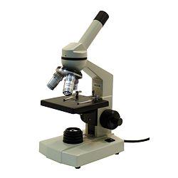
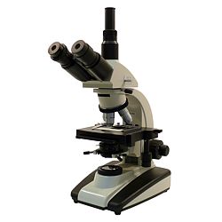
Microscopes are formed by different parts: an optical and a mechanical part. Here follows a short description of these parts:
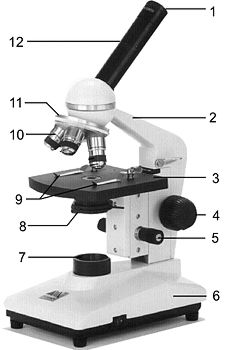
- Ocular: Are the lenses situated the closest to the beholder viewer - Arm: Situated perpendicullary to the base, they can be vertical or arched, so that they can joined to the base. - Stage: Is the part that holds the sample, it consists on two grippers and the hole from which comes the light to observe the sample - Macrometric fitting: An screw for focus and choose lenses on a quick way - Micrometric fitting. An screw for focus and choose lenses on a slow way - Stage fitting: An screw that fits the stage - Base: is the part that holds the microscope - Light souce: It is used to light the samples or items to be observed, it is isolated on the base. - Grippers: Two grippers located on the stage. Their aim is to hold the samples - Objective lens: The lense situated the closest to the sample - Revolver: Is the part that holds the set of objective lenses, it allows to change them. - Tube: Is the part were the ocular is, it holds the revolver with the objective lenses on the bottom and the oculars on the top.
Information
concerning the use of microscopes:
- Microscopes should be installed straight and safely
- Mircorscopes need to be connected to a socket
- Specimen stage on lowest level
- Object needs to be in the middle of the specimen stage
- control
illumination level with the help of the aperture
Different types of microscopes
Simple Microscopes: They just use a single magnifying glass
Composite Microscopes: They are formed by a kit of glasses, in a way that they allow to magnify the image through them. (optics microscopes).
Electronic Microscopes: They use electrons instead of visible light (fotons) to provide imag 
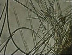
es of small items. Those icroscopes increase the speed of electrons to get a shorted wavelength and a better resolution (electrons have a wavelength rather small than visible light, so they are able to break up very small structures) It gives a magnification capacity about 500.000 magnifications compared to other optical microscopes. The images obtained are on black and white as they use electrons instead of light. The electronic spot is produced by a Wolframic cathode.
Microscopes of reflected light: These microscopes are used mainly to observe transparent and liquid preparations. They are specially used in blood analyse, cells, tests in plants. Classical reflected light microscopes have a really short working distance, under 4 mm. This is why these microscopes are suitable for very thin solutions.

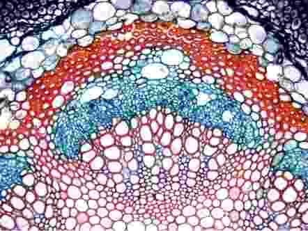
On the image above you can observe a cell that has been tinted for better observation trough microscope
Solutions are situated on the samples holder and covered with the samples cover lid. Reflected light microscopes are usually offered with a full range of magnification glasses (from 40 to more than 1000). For works of 1000 magnifications it is necessary to put a drop of oil immersion to seal the space between the sample holder and the cover lid. Images until 400 magnifications can be seen without any special technic. The magnification on reflected light microscopes can be increased by changing the oculars
Stereo Microscopes using reflected or transmitted light: These microscopes are typically used to view large objects. Their range of uses includes analysing insects, plants, coins or for testing materials. The majority of reflected light microscopes have a working distance of 40mm, making these microscopes ideal for working with large objects or for testing different materials. Normally we offer these microscopes as models with two eye pieces.
Fluorescence Microscopes: Used to reveal natural fluorescent molecules, or to reveal an added fluorescence, when detecting antibodies.

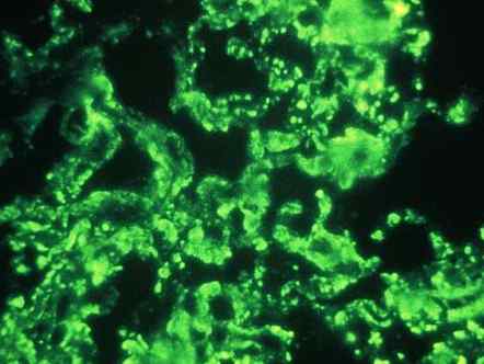
On this image you can see a sample that has not been treated, because it is fluorescent by itself. This circumstance makes tinting unnecessary
In immunity fluorescence a fluorescent dye (almost always FITC) is added to an antibody. These antibodies can be produced on a very specific way to concrete biological structures. The dyes are extremely selective, but not so intensive as in traditional secondary fluorescence.

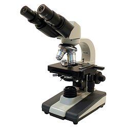
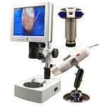
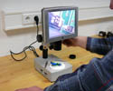
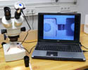
For applications that require to keep images there are trinocular microscopes. They consist on binocular microscopes with an additional tube that allows to install an USB camera that recordes images. These images can later be transferred to a PC or a laptop. It also offers the possibility to connect a microocular to the binocular microscopes. This microocular is simply set to the ocular of the microscope. This gives the opportunity to turn microscopes on video microscopes in an economical way.

Requirements to the
microscopes:
Since microscopes are used in different
areas, their equipment differs. There are normal microscopes with a
magnification from 400 to 600-fold. There are also special lighting systems
available which makes the examination of low-contrast objects possible.
Microscopes in PTB
If you wish to view or print a selection of microscopes from our catalogue, click the PDF symbol

|
|